الجوهرة المصونة
*****


المشاركات : 17164
العمـر : 36
تعاليق : مشرفة الطب والصحة
المزاج : 
الدولة : 
المهنة : 
الهواية : 
التسجيل : 10/10/2008
النقاط : 18248
التقييم : 501

 
توفيقك يارب

 |  موضوع: رد: سرطان الثدي Breast cancer الخميس نوفمبر 12, 2009 7:02 pm موضوع: رد: سرطان الثدي Breast cancer الخميس نوفمبر 12, 2009 7:02 pm | |
| Introduction to breast cancer
Breast cancer is the most common cause of cancer in women and the second most common cause of cancer death in women in the U.S. While the majority of new breast cancers are diagnosed as a result of an abnormality seen on a mammogram, a lump or change in consistency of the breast tissue can also be a warning sign of the disease. Heightened awareness of breast cancer risk in the past decades has led to an increase in the number of women undergoing mammography for screening, leading to detection of cancers in earlier stages and a resultant improvement in survival rates. Still, breast cancer is the most common cause of death in women between the ages of 45 and 55. Although breast cancer in women is a common form of cancer, male breast cancer does occur and accounts for about 1% of all cancer deaths in men.
Research has yielded much information about the causes of breast cancers, and it is now believed that genetic and/or hormonal factors are the primary risk factors for breast cancer. Staging systems have been developed to allow doctors to characterize the extent to which a particular cancer has spread and to make decisions concerning treatment options. Breast cancer treatment depends upon many factors, including thee type of cancer and the extent to which it has spread. Treatment options for breast cancer may involve surgery (removal of the cancer alone or, in some cases, mastectomy), radiation therapy, hormonal therapy, and/or chemotherapy.
With advances in screening, diagnosis, and treatment, the death rate for breast cancer has declined by about 20% over the past decade, and research is ongoing to develop even more effective screening and treatment programs.
How is the breast designed?
The breasts sit on the chest muscles that cover the ribs. Each breast is made of 15 to 20 lobes. Lobes contain many smaller lobules. Lobules contain groups of tiny glands that can produce milk. Milk flows from the lobules through thin tubes called ducts to the nipple. The nipple is in the center of a dark area of skin called the areola. Fat fills the spaces between the lobules and ducts.
The breasts also contain lymph vessels. These vessels lead to small, round organs called lymph nodes. Groups of lymph nodes are near the breast in the axilla (underarm), above the collarbone, in the chest behind the breastbone, and in many other parts of the body. The lymph nodes trap bacteria, cancer cells, or other harmful substances.

What are risk factors for breast cancer?
No one knows the exact causes of breast cancer. Doctors often cannot explain why one woman develops breast cancer and another does not. They do know that bumping, bruising, or touching the breast does not cause cancer. And breast cancer is not contagious. You cannot "catch" it from another person.
Research has shown that women with certain risk factors are more likely than others to develop breast cancer. A risk factor is something that may increase the chance of developing a disease.
Studies have found the following risk factors for breast cancer:
Age: The chance of getting breast cancer goes up as a woman gets older. Most cases of breast cancer occur in women over 60. This disease is not common before menopause.
Personal history of breast cancer: A woman who had breast cancer in one breast has an increased risk of getting cancer in her other breast.
Family history: A woman's risk of breast cancer is higher if her mother, sister, or daughter had breast cancer. The risk is higher if her family member got breast cancer before age 40. Having other relatives with breast cancer (in either her mother's or father's family) may also increase a woman's risk.
Certain breast changes: Some women have cells in the breast that look abnormal under a microscope. Having certain types of abnormal cells (atypical hyperplasia and lobular carcinoma in situ [LCIS]) increases the risk of breast cancer.
Gene changes: Changes in certain genes increase the risk of breast cancer. These genes include BRCA1, BRCA2, and others. Tests can sometimes show the presence of specific gene changes in families with many women who have had breast cancer. Health care providers may suggest ways to try to reduce the risk of breast cancer, or to improve the detection of this disease in women who have these changes in their genes.
Reproductive and menstrual history:
The older a woman is when she has her first child, the greater her chance of breast cancer.
Women who had their first menstrual period before age 12 are at an increased risk of breast cancer.
Women who went through menopause after age 55 are at an increased risk of breast cancer.
Women who never had children are at an increased risk of breast cancer.
Women who take menopausal hormone therapy with estrogen plus progestin after menopause also appear to have an increased risk of breast cancer.
Large, well-designed studies have shown no link between abortion or miscarriage and breast cancer.
Race: Breast cancer is diagnosed more often in white women than Latina, Asian, or African American women.
Radiation therapy to the chest: Women who had radiation therapy to the chest (including breasts) before age 30 are at an increased risk of breast cancer. This includes women treated with radiation for Hodgkin's lymphoma. Studies show that the younger a woman was when she received radiation treatment, the higher her risk of breast cancer later in life.
Breast density: Breast tissue may be dense or fatty. Older women whose mammograms (breast x-rays) show more dense tissue are at increased risk of breast cancer.
Taking DES (diethylstilbestrol): DES was given to some pregnant women in the United States between about 1940 and 1971. (It is no longer given to pregnant women.) Women who took DES during pregnancy may have a slightly increased risk of breast cancer. The possible effects on their daughters are under study.
Being overweight or obese after menopause: The chance of getting breast cancer after menopause is higher in women who are overweight or obese.
Lack of physical activity: Women who are physically inactive throughout life may have an increased risk of breast cancer. Being active may help reduce risk by preventing weight gain and obesity.
Drinking alcohol: Studies suggest that the more alcohol a woman drinks, the greater her risk of breast cancer.
Other possible risk factors are under study. Researchers are studying the effect of diet, physical activity, and genetics on breast cancer risk. They are also studying whether certain substances in the environment can increase the risk of breast cancer.
Many risk factors can be avoided. Others, such as family history, cannot be avoided. Women can help protect themselves by staying away from known risk factors whenever possible.
But it is also important to keep in mind that most women who have known risk factors do not get breast cancer. Also, most women with breast cancer do not have a family history of the disease. In fact, except for growing older, most women with breast cancer have no clear risk factors.
If you think you may be at risk, you should discuss this concern with your doctor. Your doctor may be able to suggest ways to reduce your risk and can plan a schedule for checkups.
What are the symptoms of breast cancer?
Common symptoms of breast cancer include:
A change in how the breast or nipple feels
A lump or thickening in or near the breast or in the underarm area
Nipple tenderness
A change in how the breast or nipple looks
A change in the size or shape of the breast
A nipple turned inward into the breast
The skin of the breast, areola, or nipple may be scaly, red, or swollen. It may have ridges or pitting so that it looks like the skin of an orange.
Nipple discharge (fluid)
Early breast cancer usually does not cause pain. Still, a woman should see her health care provider about breast pain or any other symptom that does not go away. Most often, these symptoms are not due to cancer. Other health problems may also cause them. Any woman with these symptoms should tell her doctor so that problems can be diagnosed and treated as early as possible.
How is breast cancer diagnosed?
If you have a symptom or screening test result that suggests cancer, your doctor must find out whether it is due to cancer or to some other cause. Your doctor may ask about your personal and family medical history. You may have a physical exam. Your doctor also may order a mammogram or other imaging procedure. These tests make pictures of tissues inside the breast. After the tests, your doctor may decide no other exams are needed. Your doctor may suggest that you have a follow-up exam later on. Or you may need to have a biopsy to look for cancer cells.
Clinical breast exam
Your health care provider feels each breast for lumps and looks for other problems. If you have a lump, your doctor will feel its size, shape, and texture. Your doctor will also check to see if it moves easily. Benign lumps often feel different from cancerous ones. Lumps that are soft, smooth, round, and movable are likely to be benign. A hard, oddly shaped lump that feels firmly attached within the breast is more likely to be cancer.
Diagnostic mammogram
Diagnostic mammograms are x-ray pictures of the breast. They take clearer, more detailed images of areas that look abnormal on a screening mammogram. Doctors use them to learn more about unusual breast changes, such as a lump, pain, thickening, nipple discharge, or change in breast size or shape. Diagnostic mammograms may focus on a specific area of the breast. They may involve special techniques and more views than screening mammograms.
Ultrasound
An ultrasound device sends out sound waves that people cannot hear. The waves bounce off tissues. A computer uses the echoes to create a picture. Your doctor can view these pictures on a monitor. The pictures may show whether a lump is solid or filled with fluid. A cyst is a fluid-filled sac. Cysts are not cancer. But a solid mass may be cancer. After the test, your doctor can store the pictures on video or print them out. This exam may be used along with a mammogram.
Magnetic resonance imaging
Magnetic resonance imaging (MRI) uses a powerful magnet linked to a computer. MRI makes detailed pictures of breast tissue. Your doctor can view these pictures on a monitor or print them on film. MRI may be used along with a mammogram.
Biopsy
Your doctor may refer you to a surgeon or breast disease specialist for a biopsy. Fluid or tissue is removed from your breast to help find out if there is cancer.
Some suspicious areas can be seen on a mammogram but cannot be felt during a clinical breast exam. Doctors can use imaging procedures to help see the area and remove tissue. Such procedures include ultrasound-guided, needle-localized, or stereotactic biopsy.
Doctors can remove tissue from the breast in different ways:
Fine-needle aspiration: Your doctor uses a thin needle to remove fluid from a breast lump. If the fluid appears to contain cells, a pathologist at a lab checks them for cancer with a microscope. If the fluid is clear, it may not need to be checked by a lab.
Core biopsy: Your doctor uses a thick needle to remove breast tissue. A pathologist checks for cancer cells. This procedure is also called a needle biopsy.
Surgical biopsy: Your surgeon removes a sample of tissue. A pathologist checks the tissue for cancer cells.
An incisional biopsy takes a sample of a lump or abnormal area.
An excisional biopsy takes the entire lump or area.
If cancer cells are found, the pathologist can tell what kind of cancer it is. The most common type of breast cancer is ductal carcinoma. Abnormal cells are found in the lining of the ducts. Lobular carcinoma is another type. Abnormal cells are found in the lobules.
What is the screening process for breast cancer?
Screening for breast cancer before there are symptoms can be important. Screening can help doctors find and treat cancer early. Treatment is more likely to work well when cancer is found early.
Your doctor may suggest the following screening tests for breast cancer:
Screening mammogram
Clinical breast exam
Breast self-exam
You should ask your doctor about when to start and how often to check for breast cancer.
Screening mammogram
To find breast cancer early, NCI recommends that:
Women in their 40s and older should have mammograms every 1 to 2 years. A mammogram is a picture of the breast made with x-rays.
Women who are younger than 40 and have risk factors for breast cancer should ask their health care provider whether to have mammograms and how often to have them.
Mammograms can often show a breast lump before it can be felt. They also can show a cluster of tiny specks of calcium. These specks are called microcalcifications. Lumps or specks can be from cancer, precancerous cells, or other conditions. Further tests are needed to find out if abnormal cells are present.
If an abnormal area shows up on your mammogram, you may need to have more x-rays. You also may need a biopsy. A biopsy is the only way to tell for sure if cancer is present.
Mammograms are the best tool doctors have to find breast cancer early. However, mammograms are not perfect:
A mammogram may miss some cancers. (The result is called a "false negative.")
A mammogram may show things that turn out not to be cancer. (The result is called a "false positive.")
Some fast-growing tumors may grow large or spread to other parts of the body before a mammogram detects them.
Mammograms (as well as dental x-rays, and other routine x-rays) use very small doses of radiation. The risk of any harm is very slight, but repeated x-rays could cause problems. The benefits nearly always outweigh the risk. You should talk with your health care provider about the need for each x-ray. You should also ask for shields to protect parts of your body that are not in the picture.
Clinical breast exam
During a clinical breast exam, your health care provider checks your breasts. You may be asked to raise your arms over your head, let them hang by your sides, or press your hands against your hips.
Your health care provider looks for differences in size or shape between your breasts. The skin of your breasts is checked for a rash, dimpling, or other abnormal signs. Your nipples may be squeezed to check for fluid.
Using the pads of the fingers to feel for lumps, your health care provider checks your entire breast, underarm, and collarbone area. A lump is generally the size of a pea before anyone can feel it. The exam is done on one side, then the other. Your health care provider checks the lymph nodes near the breast to see if they are enlarged.
A thorough clinical breast exam may take about 10 minutes.
Breast self-exam
You may perform monthly breast self-exams to check for any changes in your breasts. It is important to remember that changes can occur because of aging, your menstrual cycle, pregnancy, menopause, or taking birth control pills or other hormones. It is normal for breasts to feel a little lumpy and uneven. Also, it is common for your breasts to be swollen and tender right before or during your menstrual period.
You should contact your health care provider if you notice any unusual changes in your breasts.
Breast self-exams cannot replace regular screening mammograms and clinical breast exams. Studies have not shown that breast self-exams alone reduce the number of deaths from breast cancer.
How is breast cancer staging determined?
To plan your treatment, your doctor needs to know the extent (stage) of the disease. The stage is based on the size of the tumor and whether the cancer has spread. Staging may involve x-rays and lab tests. These tests can show whether the cancer has spread and, if so, to what parts of your body. When breast cancer spreads, cancer cells are often found in lymph nodes under the arm (axillary lymph nodes). The stage often is not known until after surgery to remove the tumor in your breast and the lymph nodes under your arm.
These are the stages of breast cancer:
Stage 0 is carcinoma in situ.
Lobular carcinoma in situ (LCIS): Abnormal cells are in the lining of a lobule. LCIS seldom becomes invasive cancer. However, having LCIS in one breast increases the risk of cancer for both breasts.
Ductal carcinoma in situ (DCIS): Abnormal cells are in the lining of a duct. DCIS is also called intraductal carcinoma. The abnormal cells have not spread outside the duct. They have not invaded the nearby breast tissue. DCIS sometimes becomes invasive cancer if not treated.
Stage I is an early stage of invasive breast cancer. The tumor is no more than 2 centimeters (three-quarters of an inch) across. Cancer cells have not spread beyond the breast.
Stage II is one of the following:
The tumor in the breast is no more than 2 centimeters (three-quarters of an inch) across. The cancer has spread to the lymph nodes under the arm.
The tumor is between 2 and 5 centimeters (three-quarters of an inch to 2 inches). The cancer may have spread to the lymph nodes under the arm.
The tumor is larger than 5 centimeters (2 inches). The cancer has not spread to the lymph nodes under the arm.
Stage III is locally advanced cancer. It is divided into Stage IIIA, IIIB, and IIIC.
Stage IIIA is one of the following:
The tumor in the breast is smaller than 5 centimeters (2 inches). The cancer has spread to underarm lymph nodes that are attached to each other or to other structures. Or the cancer may have spread to lymph nodes behind the breastbone.
The tumor is more than 5 centimeters across. The cancer has spread to the underarm lymph nodes that are either alone or attached to each other or to other structures. Or the cancer may have spread to lymph nodes behind the breastbone.
Stage IIIB is a tumor of any size that has grown into the chest wall or the skin of the breast. It may be associated with swelling of the breast or with nodules (lumps) in the breast skin.
The cancer may have spread to lymph nodes under the arm.
The cancer may have spread to underarm lymph nodes that are attached to each other or other structures. Or the cancer may have spread to lymph nodes behind the breastbone.
Inflammatory breast cancer is a rare type of breast cancer. The breast looks red and swollen because cancer cells block the lymph vessels in the skin of the breast. When a doctor diagnoses inflammatory breast cancer, it is at least Stage IIIB, but it could be more advanced.
Stage IIIC is a tumor of any size. It has spread in one of the following ways:
The cancer has spread to the lymph nodes behind the breastbone and under the arm.
The cancer has spread to the lymph nodes under or above the collarbone.
Stage IV is distant metastatic cancer. The cancer has spread to other parts of the body.
Recurrent cancer is cancer that has come back (recurred) after a period of time when it could not be detected. It may recur locally in the breast or chest wall. Or it may recur in any other part of the body, such as the bone, liver, or lungs.
What are the methods for treating breast cancer?
Many women with breast cancer want to take an active part in making decisions about their medical care. It is natural to want to learn all you can about your disease and treatment choices. Knowing more about breast cancer helps many women cope.
Shock and stress after the diagnosis can make it hard to think of everything you want to ask your doctor. It often helps to make a list of questions before an appointment. To help remember what the doctor says, you may take notes or ask whether you may use a tape recorder. You may also want to have a family member or friend with you when you talk to the doctor - to take part in the discussion, to take notes, or just to listen. You do not need to ask all your questions at once. You will have other chances to ask your doctor or nurse to explain things that are not clear and to ask for more details.
Your doctor may refer you to a specialist, or you may ask for a referral. Specialists who treat breast cancer include surgeons, medical oncologists, and radiation oncologists. You also may be referred to a plastic surgeon.
Getting a second opinion
Before starting treatment, you might want a second opinion about your diagnosis and treatment plan. Many insurance companies cover a second opinion if you or your doctor requests it. It may take some time and effort to gather medical records and arrange to see another doctor. You may have to gather your mammogram films, biopsy slides, pathology report, and proposed treatment plan. Usually it is not a problem to take several weeks to get a second opinion. In most cases, the delay in starting treatment will not make treatment less effective. To make sure, you should discuss this delay with your doctor. Some women with breast cancer need treatment right away.
There are a number of ways to find a doctor for a second opinion:
Your doctor may refer you to one or more specialists. At cancer centers, several specialists often work together as a team.
NCI's Cancer Information Service, at 1-800-4-CANCER, can tell you about nearby treatment centers. Information Specialists also can provide online assistance through LiveHelp at http://www.cancer.gov/cis.
A local or state medical society, a nearby hospital, or a medical school can usually provide the names of specialists.
The American Board of Medical Specialties (ABMS) has a list of doctors who have had training and passed exams in their specialty. You can find this list in the Official ABMS Directory of Board Certified Medical Specialists. This Directory is in most public libraries. Also, ABMS offers this information at http://www.abms.org. (Click on "Who's Certified.")
NCI provides a helpful fact sheet called "How To Find a Doctor or Treatment Facility If You Have Cancer."
Treatment methods
Women with breast cancer have many treatment options. These include surgery, radiation therapy, chemotherapy, hormone therapy, and biological therapy. These options are described below. Many women receive more than one type of treatment.
The choice of treatment depends mainly on the stage of the disease. Treatment options by stage are described below.
Your doctor can describe your treatment choices and the expected results. You may want to know how treatment may change your normal activities. You may want to know how you will look during and after treatment. You and your doctor can work together to develop a treatment plan that reflects your medical needs and personal values.
Cancer treatment is either local therapy or systemic therapy:
Local therapy: Surgery and radiation therapy are local treatments. They remove or destroy cancer in the breast. When breast cancer has spread to other parts of the body, local therapy may be used to control the disease in those specific areas.
Systemic therapy: Chemotherapy, hormone therapy, and biological therapy are systemic treatments. They enter the bloodstream and destroy or control cancer throughout the body. Some women with breast cancer have systemic therapy to shrink the tumor before surgery or radiation. Others have systemic therapy after surgery and/or radiation to prevent the cancer from coming back. Systemic treatments also are used for cancer that has spread.
Because cancer treatments often damage healthy cells and tissues, side effects are common. Side effects depend mainly on the type and extent of the treatment. Side effects may not be the same for each woman, and they may change from one treatment session to the next.
Before treatment starts, your health care team will explain possible side effects and suggest ways to help you manage them.
At any stage of disease, supportive care is available to control pain and other symptoms, to relieve the side effects of treatment, and to ease emotional concerns.
You may want to talk to your doctor about taking part in a clinical trial, a research study of new treatment methods.
Surgery
Surgery is the most common treatment for breast cancer. There are several types of surgery. Your doctor can explain each type, discuss and compare the benefits and risks, and describe how each will change the way you look:
Breast-sparing surgery: An operation to remove the cancer but not the breast is breast-sparing surgery. It is also called breast-conserving surgery, lumpectomy, segmental mastectomy, and partial mastectomy. Sometimes an excisional biopsy serves as a lumpectomy because the surgeon removes the whole lump.
The surgeon often removes the underarm lymph nodes as well. A separate incision is made. This procedure is called an axillary lymph node dissection. It shows whether cancer cells have entered the lymphatic system.
After breast-sparing surgery, most women receive radiation therapy to the breast. This treatment destroys cancer cells that may remain in the breast.
Mastectomy: An operation to remove the breast (or as much of the breast tissue as possible) is a mastectomy. In most cases, the surgeon also removes lymph nodes under the arm. Some women have radiation therapy after surgery.
Studies have found equal survival rates for breast-sparing surgery (with radiation therapy) and mastectomy for Stage I and Stage II breast cancer.
Sentinel lymph node biopsy is a new method of checking for cancer cells in the lymph nodes. A surgeon removes fewer lymph nodes, which causes fewer side effects. (If the doctor finds cancer cells in the axillary lymph nodes, an axillary lymph node dissection usually is done.)
In breast-sparing surgery, the surgeon removes the tumor in the breast and some tissue around it. The surgeon may also remove lymph nodes under the arm. The surgeon sometimes removes some of the lining over the chest muscles below the tumor.
In total (simple) mastectomy, the surgeon removes the whole breast. Some lymph nodes under the arm may also be removed.
In modified radical mastectomy, the surgeon removes the whole breast, and most or all of the lymph nodes under the arm. Often, the lining over the chest muscles is removed. A small chest muscle also may be taken out to make it easier to remove the lymph nodes.
You may choose to have breast reconstruction. This is plastic surgery to rebuild the shape of the breast. It may be done at the same time as a mastectomy or later. If you are considering reconstruction, you may wish to talk with a plastic surgeon before having a mastectomy. More information is in the "Breast Reconstruction" section.
The time it takes to heal after surgery is different for each woman. Surgery causes pain and tenderness. Medicine can help control the pain. Before surgery, you should discuss the plan for pain relief with your doctor or nurse. After surgery, your doctor can adjust the plan if you need more relief. Any kind of surgery also carries a risk of infection, bleeding, or other problems. You should tell your health care provider right away if you develop any problems.
You may feel off balance if you've had one or both breasts removed. You may feel more off balance if you have large breasts. This imbalance can cause discomfort in your neck and back. Also, the skin where your breast was removed may feel tight. Your arm and shoulder muscles may feel stiff and weak. These problems usually go away. The doctor, nurse, or physical therapist can suggest exercises to help you regain movement and strength in your arm and shoulder. Exercise can also reduce stiffness and pain. You may be able to begin gentle exercises within days of surgery.
Because nerves may be injured or cut during surgery, you may have numbness and tingling in your chest, underarm, shoulder, and upper arm. These feelings usually go away within a few weeks or months. But for some women, numbness does not go away.
Removing the lymph nodes under the arm slows the flow of lymph fluid. The fluid may build up in your arm and hand and cause swelling. This swelling is lymphedema. Lymphedema can develop right after surgery or months to years later.
You will need to protect your arm and hand on the treated side for the rest of your life:
Avoid wearing tight clothing or jewelry on your affected arm
Carry your purse or luggage with the other arm
Use an electric razor to avoid cuts when shaving under your arm
Have shots, blood tests, and blood pressure measurements on the other arm
Wear gloves to protect your hands when gardening and when using strong detergents
Have careful manicures and avoid cutting your cuticles
Avoid burns or sunburns to your affected arm and hand
You should ask your doctor how to handle any cuts, insect bites, sunburn, or other injuries to your arm or hand. Also, you should contact the doctor if your arm or hand is injured, swells, or becomes red and warm.
If lymphedema occurs, the doctor may suggest raising your arm above your heart whenever you can. The doctor may show you hand and arm exercises. Some women with lymphedema wear an elastic sleeve to improve lymph circulation. Medication, manual lymph drainage (massage), or use of a machine that gently compresses the arm may also help. You may be referred to a physical therapist or another specialist.
Radiation therapy
Radiation therapy (also called radiotherapy) uses high-energy rays to kill cancer cells. Most women receive radiation therapy after breast-sparing surgery. Some women receive radiation therapy after a mastectomy. Treatment depends on the size of the tumor and other factors. The radiation destroys breast cancer cells that may remain in the area.
Some women have radiation therapy before surgery to destroy cancer cells and shrink the tumor. Doctors use this approach when the tumor is large or may be hard to remove. Some women also have chemotherapy or hormone therapy before surgery.
Doctors use two types of radiation therapy to treat breast cancer. Some women receive both types:
External radiation: The radiation comes from a large machine outside the body. Most women go to a hospital or clinic for treatment. Treatments are usually 5 days a week for several weeks.
Internal radiation (implant radiation): Thin plastic tubes (implants) that hold a radioactive substance are put directly in the breast. The implants stay in place for several days. A woman stays in the hospital while she has implants. Doctors remove the implants before she goes home.
Side effects depend mainly on the dose and type of radiation and the part of your body that is treated.
It is common for the skin in the treated area to become red, dry, tender, and itchy. Your breast may feel heavy and tight. These problems will go away over time. Toward the end of treatment, your skin may become moist and "weepy." Exposing this area to air as much as possible can help the skin heal.
Bras and some other types of clothing may rub your skin and cause soreness. You may want to wear loose-fitting cotton clothes during this time. Gentle skin care also is important. You should check with your doctor before using any deodorants, lotions, or creams on the treated area. These effects of radiation therapy on the skin will go away. The area gradually heals once treatment is over. However, there may be a lasting change in the color of your skin.
You are likely to become very tired during radiation therapy, especially in the later weeks of treatment. Resting is important, but doctors usually advise patients to try to stay as active as they can.
Although the side effects of radiation therapy can be distressing, your doctor can usually relieve them.
Chemotherapy
Chemotherapy uses anticancer drugs to kill cancer cells. Chemotherapy for breast cancer is usually a combination of drugs. The drugs may be given as a pill or by injection into a vein (IV). Either way, the drugs enter the bloodstream and travel throughout the body.
Women with breast cancer can have chemotherapy in an outpatient part of the hospital, at the doctor's office, or at home. Some women need to stay in the hospital during treatment.
Side effects depend mainly on the specific drugs and the dose. The drugs affect cancer cells and other cells that divide rapidly:
Blood cells: These cells fight infection, help your blood to clot, and carry oxygen to all parts of the body. When drugs affect your blood cells, you are more likely to get infections, bruise or bleed easily, and feel very weak and tired. Years after chemotherapy, some women have developed leukemia (cancer of the blood cells).
Cells in hair roots: Chemotherapy can cause hair loss. Your hair will grow back, but it may be somewhat different in color and texture.
Cells that line the digestive tract: Chemotherapy can cause poor appetite, nausea and vomiting, diarrhea, or mouth and lip sores.
Your doctor can suggest ways to control many of these side effects.
Some drugs used for breast cancer can cause tingling or numbness in the hands or feet. This problem usually goes away after treatment is over. Other problems may not go away. In some women, the drugs used for breast cancer may weaken the heart.
Some anticancer drugs can damage the ovaries. The ovaries may stop making hormones. You may have symptoms of menopause. The symptoms include hot flashes and vaginal dryness. Your menstrual periods may no longer be regular or may stop. Some women become infertile (unable to become pregnant). For women over the age of 35, infertility is likely to be permanent.
On the other hand, you may remain fertile during chemotherapy and be able to become pregnant. The effects of chemotherapy on an unborn child are not known. You should talk to your doctor about birth control before treatment begins.
Hormone therapy
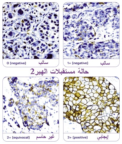
Some breast tumors need hormones to grow. Hormone therapy keeps cancer cells from getting or using the natural hormones they need. These hormones are estrogen and progesterone. Lab tests can show if a breast tumor has hormone receptors. If you have this kind of tumor, you may have hormone therapy.
This treatment uses drugs or surgery:
Drugs: Your doctor may suggest a drug that can block the natural hormone. One drug is tamoxifen, which blocks estrogen. Another type of drug prevents the body from making the female hormone estradiol. Estradiol is a form of estrogen. This type of drug is an aromatase inhibitor. If you have not gone through menopause, your doctor may give you a drug that stops the ovaries from making estrogen.
Surgery: If you have not gone through menopause, you may have surgery to remove your ovaries. The ovaries are the main source of the body's estrogen. A woman who has gone through menopause does not need surgery. (The ovaries produce less estrogen after menopause.)
The side effects of hormone therapy depend largely on the specific drug or type of treatment. Tamoxifen is the most common hormone treatment. In general, the side effects of tamoxifen are similar to some of the symptoms of menopause. The most common are hot flashes and vaginal discharge. Other side effects are irregular menstrual periods, headaches, fatigue, nausea, vomiting, vaginal dryness or itching, irritation of the skin around the vagina, and skin rash. Not all women who take tamoxifen have side effects.
It is possible to become pregnant when taking tamoxifen. Tamoxifen may harm the unborn baby. If you are still menstruating, you should discuss birth control methods with your doctor.
Serious side effects of tamoxifen are rare. However, it can cause blood clots in the veins. Blood clots form most often in the legs and in the lungs. Women have a slight increase in their risk of stroke.
Tamoxifen can cause cancer of the uterus. Your doctor should perform regular pelvic exams. You should tell your doctor about any unusual vaginal bleeding between exams.
When the ovaries are removed, menopause occurs at once. The side effects are often more severe than those caused by natural menopause. Your health care provider can suggest ways to cope with these side effects. | |
|